CLADOENDESIS OF EPHEMEROPTERA
|
CLADOENDESIS OF EPHEMEROPTERA |
||||
| zzz | ||||
|
|
|
Kimminsula/g1
(Panephemeroptera![]() Euephemeroptera
Euephemeroptera![]() Euplectoptera
Euplectoptera![]() Anteritorna
Anteritorna![]() Bidentiseta
Bidentiseta![]() Furcatergaliae
Furcatergaliae![]()
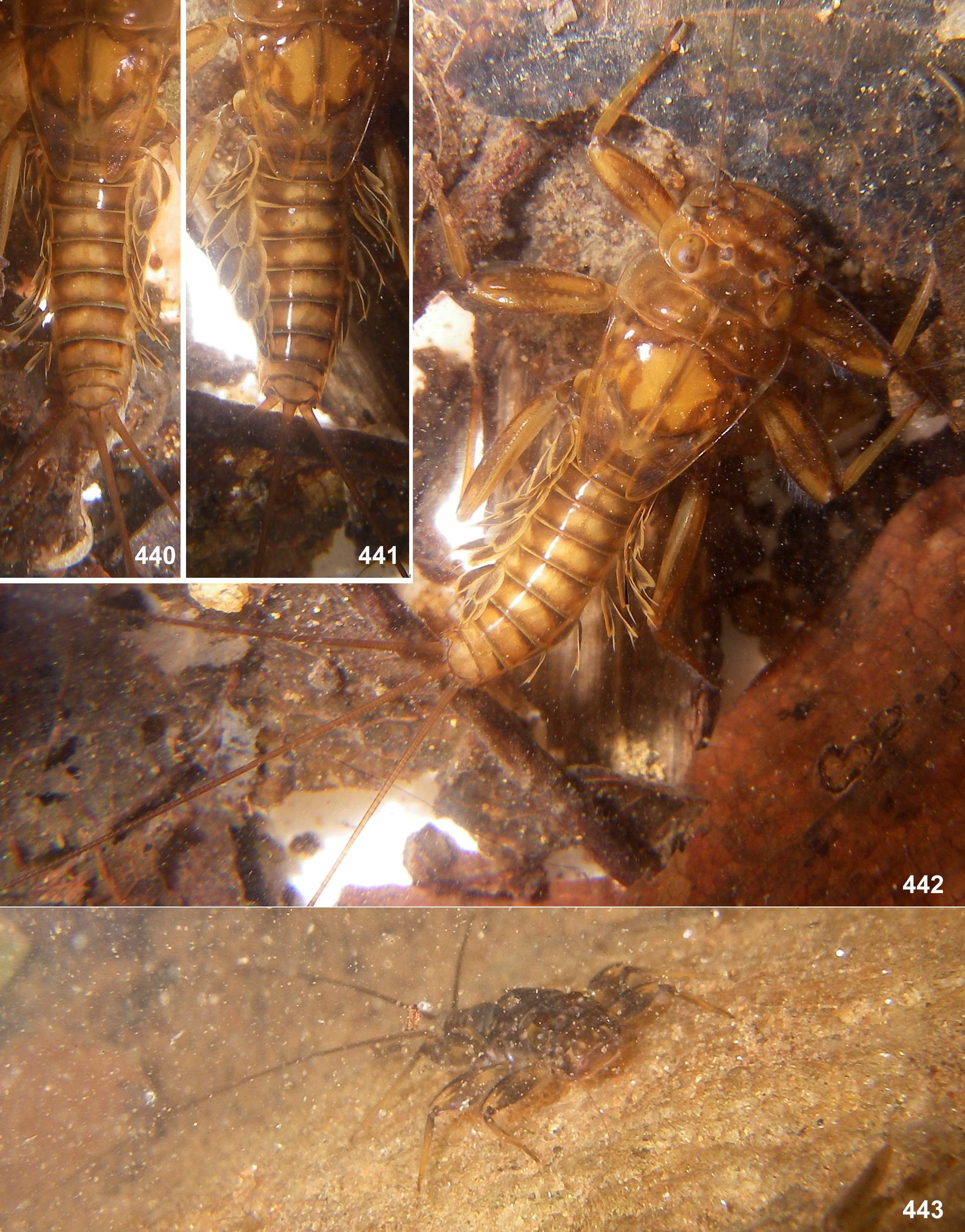
Leptophlebia/fg1![]() Atalophleboadentata
Atalophleboadentata![]() Atalophlebopectinata
Atalophlebopectinata![]() Atalophleboculata
Atalophleboculata![]() Atalophlebomaxillata
Atalophlebomaxillata![]() Atalophlebolinguata
Atalophlebolinguata![]() ... Kimminsula/g1)
... Kimminsula/g1)
Nomen hierarchicum:
![]() Kimminsula/g1 [g:1970]
(incl. Ceylonula, Hubbardula, Petersula, Ghatula)
Kimminsula/g1 [g:1970]
(incl. Ceylonula, Hubbardula, Petersula, Ghatula)
In circumscription fits:
— Kimminsula-complex: Kluge & Vasanth & Balasubrtamanian & Sivaramakrishnan 2022
References.
Kluge & Vasanth & Balasubrtamanian & Sivaramakrishnan 2022: ![]()
![]()
|
Characters of Kimminsula/g1 of unclear phylogenetic status. () All imaginal and subimaginal claws are pointed. Non-unique apomorphy (see Index of characters [2.2.85]). (1) General appearance. Larva has flattened body, widened head and enlarged legs with broad femora and claws bent perpendicular to the leg flatness (Figs 202, 440–443). Superficially, they resemble larvae of Heptageniidae, but in contrast to them have no frontal shield; instead, the wide dorsal head surface contains surfaces of closely pressed together clypeus, labrum and mandibles (Fig. 235) (the same in other Atalophlebomaxillata). Initially such larval structure is an adaptation for inhabiting stone surfaces in rapidly running water (Fig. 219), but larvae of Ceylonula femoralis inhabit soft substrates in stagnant water (Fig. 220). (2) Fronto-clypeal incisions. Lateral margins of the clypeus are separated from lateral margins of the frons by a pair of sharp incisions leading into the anterior tentorial pits; within these incisions, the margins of the frons overlap the margins of the clypeus dorsally (Figs 3, 14, 68, 105, 235, 335, 389, 444, 485, 552–553, 616). This differs from many other Atalophlebolinguata, where the frons and the clypeus have an integral dorsal surface, being separated only by a pair of smooth concavities of the lateral margins (Figs 5–6) (see below, discussion of systematic position of the Kimminsula-complex). (3) Labrum. As in most other Atalophlebopectinata, the dorsal surface of the labrum bears the posterior transverse setal row and the anterior transverse setal row; in the Kimminsula-complex both rows stretch nearly across the labrum width; each row is dense, either regular, i.e. with one seta per section (Figs 16, 73, 232: anter.r, 488, 619), or has the form of a strip with a few setae per section (Figs 102, 232: poster.r, 617). In all species of Kimminsula-complex, the anterior margin of labrum is hooded, i.e. the initial anterior margin is bent ventrally, so that in dorsal view the posterior transverse setal row appears to be located on the visible anterior margin or close to it, while the anterior transverse setal row is often hidden ventrally (Fig. 101). Setae of the true ventral side of labrum are situated irregularly (i.e. not forming rows), and form a distinctly outlined transverse ventro-anterior strip close to the anterior margin (Figs 101, 103). In all species except Ghatula rufa gen. sp. n., labrum is wider than clypeus. Shape of the median incision is variable: either shallow with more or less developed denticles on the initial anterior margin (in the Ceylonese taxa Ceylonula gen. n., Kimminsula and Hubbardula gen. n., Figs 233, 487, 619), or cleft-like and lacking denticles (in the Indian taxa Ghatula gen. n. and Petersula, Figs 17, 103). Petersula differs from other taxa by peculiar labral depressors [see below, Petersula (2)] (4) Mandibles. As in majority of Atalophlebolinguata, mandibles are articulated at ventral side of the head capsule some distance from its lateral margins (Fig. 235); mandibles are flattened, with outer margins convex and projected laterally. The outer margin of mandible bears the distal tuft of setae, which occupies an area adjacent to the base of the incisor, and the middle tuft of setae which is more compact and located more laterally (Figs 22–23, 75–76, 229, 563, 620). Among members of the Kimminsula-complex, the middle tuft is absent only in Petersula (Figs 145–146). The same in some other leptophlebiid taxa (Kluge 2020). (5) Maxillae. Maxilla is wide. The subapical ventral row of comb-like setae (peculiar for Leptophlebiidae) has more or less expressed curvature, which divides it into two portions: a lateral portion consisting of setae with smaller sockets, and a median portions consisting of setae with larger sockets (length of all setae is equal). In the Ceylonese taxa, i.e. Ceylonula gen. n., Kimminsula and Hubbardula gen. n., the lateral portion is very long and the median portion is short, consisting of 5–10 setae (Figs 332–334). In the Indian taxa, i.e. Ghatula gen. n. and Petersula, the whole subapical ventral setal row is shorter, and its both portions are subequal in length (Figs 112–113) (the same in many other leptophlebiid taxa). (6) Maxillary palps. Maxillary palp bears long setae on outer sides of the 2nd and 3rd segments, dense setae on ventral side and on apex of the 3rd segment. The inner side of the 3rd segment bears a regular row of setae, whose size and number vary individually and do not provide taxonomic characters within the Kimminsula-complex. The inner side of the 2nd segment bears longer setae which are either concentrated at its apex (in both species of Petersula, Fig. 111) or form a more or less long longitudinal row (in other species, Figs 19, 236, 334, 493, 566, 623). (7) Labium. Shape and setation of labium is uniform in all representatives (Figs 26, 77, 116–118, 241, 492, 567, 624–625). Submentum is bare without prominent setae: either without visible setae at all (Figs 116, 567, 625), or with very small occasional setae. The glossae are not large, not projected from paraglossae neither ventrally, nor dorsally; long setae of their ventral side are situated densely and irregularly (Fig. 625); long setae on the dorsal side of glossa form irregular row or strip of a horseshoe shape (Fig. 242, 624) [as in Thaulodes (Kuge 2020: fig. 20)]. (8) Labial palps. The last (3rd) segment of labial palp is shorter and narrower than others and apically blunt. A small, regular row of stout claw-like setae crosses apex of this segment. In Petersula, these claw-like setae are relatively long (Fig. 117); in other taxa they are very short, with length about twice exceeding width at base (Figs 25, 240, 492, 567, 626). The same condition occurs in most other Atalophleboculata (see discussion about systematic position below). (9) Larval thorax. The pronotum is nearly rectangular, with rounded antero-lateral angles and a free anterior margin (i.e. transverse margin between antero-lateral angles and the neck membrane); a transverse ridge runs parallel to the free anterior margin; humeral setae are located between the free anterior margin and the ridge, being absent in other places (Figs 9, 91, 95, 228, 324, 391, 481, 556, 610) (the same in many other Atalophlebolinguata). (10) Larval femora. Femora are widened, so the groove on inner side (into which the tibia can be partly inserted) is bordered with prominent flanges (called here the inner-anterior flange and the inner-posterior flange); at least on the middle femur, the inner-anterior flange is expanded in its distal part, so the femur appears widest in distal part (Fig. 28); the forefemur has similar shape in all species except Ghatula rufa gen sp. n., in which the inner-anterior flange of forefemur is reduced, being narrower than the inner-posterior flange (Fig. 27); the hind femur retains the usual elongate-ellipsoid shape, being widest in the middle (Fig. 29). The forefemur is not widened proximally (in contrast to many other Leptophlebiidae), but the cuticle of its anterior surface bears the proximal blank peculiar for mayfly larvae whose forefemur is proximally wider (Kluge 2020: p. 15). The proximal blank of foreleg differs from blanks occurring on the middle and hind legs (Fig 27, 119, 243, 336, 392, 449, 496, 571, 626). The anterior surface of each femur bears more or less numerous, irregularly situated, stout setae, which can be either blunt (Fig. 494), or pointed (Fig. 495), or variable. On the inner side of the femur the stout setae form a more or less regular row running by margin of the inner-anterior flange. On middle and hind femora (but never on forefemur) the stout setae form a regular, arched, transverse row situated near the femur base (Figs 28–29, 79–80, 120–121, 245, 337–338, 497–498, 572–573, 627). On outer side of each femur the stout setae are enlarged and form two more or less regular rows (which can be called the outer-anterior row and the outer-posterior row), between which the outer strip of thin hairs is located; hairs of this strip are situated densely and irregularly (Fig. 629). Hairs of the outer strip are generally longer and/or denser on the fore- and the middle femora, than on the hind femur (Figs 27–29, 78–80, 119–121, 336–338, 392–394, 449–451, 496–498, 571–573, 612–614); in Ceylonula gen. n. they are completely absent on the hind femur (Fig. 245). The posterior side of the hind femur bears curved setae—these are small, pointed, usually pectinate setae, curved toward the inner margin of the femur and forming an irregular strip along inner margin of the femur (Figs 36, 123, 247). The curved setae are probably initial for Leptophlebiidae; other features of femur setation, especially the transverse proximal rows on middle and hind femora, distinguish the Kimminsula-complex from many other Leptophlebiidae (see below, discussion about systematic position of the Kimminsula-complex). (11) Larval tibiae. Patella-tibial suture, which initially for mayflies is absent on the forelegs and present on the middle and the hind legs (Kluge 2004), retains this condition in Ghatula gen. n., Ceylonula gen. n. and Hubbardula gen.n.; it is differently modified in Petersula and in Kimminsula [see below, Petersula (7) and Kimminsula (7)].
As in most other Leptophlebiidae, tibiae of three leg pairs are differentiated: the pointed pectinate setae of inner side are preferably developed on the foreleg, and the stout setae of outer side are preferably developed on the hind leg. The hind tibia differs from other tibiae by the presence of stout setae located on all sides, forming two longitudinal rows on outer side of the tibia—the outer-posterior and the outer-anterior ones (Figs 258, 504–505, 639) and forming a transverse row on posterior side at the tibia apex (Figs 35, 259). As in many other Leptophlebiidae, the outer side of each tibia bears a longitudinal row of hairs (long and slender setae). In Ghatula gen. n. and Ceylonula gen. n. this row of hairs is single, but Petersula, Kimminsula and Hubbardula gen. n. have a second row located more anteriorly. Each of these rows is either regular, i.e. with one seta per section (Figs 124–125, 342, 501), or has a form of strip with a few setae per section (Figs 576, 639). On the hind tibia, which has two rows of stout setae on the outer side, the initial outer row of hairs is located between them, and the additional anterior row of hairs is located anteriad of the outer-anterior row of stout setae (Fig. 639). (12) Larval tarsus. The tarsus of each leg pair bears hairs on the outer side similar to hairs on the tibia; these hairs are longer and denser on the hind leg. Stout setae on inner sides of tarsi are differentiated as follows: on the fore- and middle legs they are small and few, can be absent; on the hind leg stout setae always form a longitudinal row at least on the distal part of the tarsus; in proximal part of the row setae are sparse and small, but they become denser, larger and hooked toward apex of the tarsus (Fig. 35). Sometimes inner side of the foretarsus bears several stout, pointed, pectinate setae of the same structure as the setae on inner side of the tibia of the same leg; such setae occur in Ceylonula femoralis (Figs 251, 253) and some others. (13) Larval claw. Claw is bent perpendicular to the leg flatness and distinctly divided into the small articulatory portion and the large rigid portion (Figs 129, 506) (Kluge 2020). (14) Larval abdomen. The surface of terga and sterna is always smooth, without prominent relief or significant setae. Posterior margin of each abdominal tergum bears a row of sharply pointed denticles greatly varying in size; medium size of denticles is either subequal on all terga I–X (Figs 38–41), or significantly differs on different terga. Posterior margin of the 10th tergum always has shallow median concavity (Figs 41, 398, 586, 631). Posterior margins of all sterna I–IX are always smooth, without denticles (Fig. 94). Caudalii are about 2–3 times longer than the body; all 3 caudalii (the cerci and the paracercus) are equally developed both in Ceylonula femoralis (whose paracercus is vestigial in the winged stages, Figs 269–271) and in other species (where the paracercus is equal to the cerci in all stages); posterior margin of each segment of cercus and paracercus bears whorls of pointed denticles and setae in all species (Figs 41–42). (15) Tergalii. Tergalii always retain 2 lamellae (initial for Leptophlebiidae); tergalii of all pairs have similar structure, with both lamellae similar. Tergalii make rhythmic undulate movements with the wave going from front to back; either all pairs of tergalii participate equally in respiration (Figs 440–442) or tergalii I do not participate in respiratory movement (Fig. 202); the same in some other taxa (see Index of characters [1.3.33]). In other respects, structure of tergalii significantly differs in different taxa [see below, Ghatula (), Petersula/g1 (), Kimminsula/g3 () and Hubbardula ()]. All 7 pairs of tergalii are retained in Petersula, Ceylonula gen. n., Kimminsula and Hubbardula gen. n., while Ghatula gen. n. has no tergalii of 7th pair, thus retains only 6 pairs of tergalii. (16) Forewings. Furcation of MA is symmetrical or slightly asymmetrical. Furcation of MP is more proximal than furcation of RS and is sharply asymmetrical: vein MP2 is attached at base to veins MP1 and CuA either by equally oblique crossveins (Fig. 411), or the crossvein connecting MP2 with MP1 is more oblique and may be interpreted as asymmetric arising of MP2 from MP1 (Figs 51, 68, 151, 156, 279, 453, 539, 670); this difference may be individual (Figs 216, 218). Cubital field has 2 intercalaries between Cu1 and Cu2; in Kimminsula latifolia sp. n. a third, additional intercalary is present between them (Fig. 581). Crossveins are numerous; some crossveins reach posterior margin of the wing between ends of the longitudinal and the intercalary veins. Crossveins in the area of pterostigma are numerous, complete and non-anastomosed; in Ceylonula femoralis the costal field is wider than the subcostal field, and the pterostigmatic crossveins are nearly perpendicular to Sc (Fig. 279); in other taxa the costal field is not wider than the subcostal field, and the pterostigmatic crossveins vary from sharply oblique (Fig. 414) to perpendicular to Sc (Figs 670). (17) Hind wings. Forewing has well expressed tornus, and the hind wing is relatively large, about as long as the basi-tornal margin of the forewing. Costal projection of the hind wing is blunt and slightly projected; Sc is continued far distad of the costal projection (Figs 52, 153, 157–158, 217, 278, 365, 412, 454, 540, 671). The vein MA is non-bifurcate as in all other Furcatergaliae (see Kluge 2004); other veins are either fully developed (Figs 157–158), or MP is non-bifurcate (in Ghatula gen. n., Fig. 52). (18) Claws of winged stages. On each leg of imago and subimago, both claws are pointed and hooked (Figs 289, 676) (see Index of characters [2.2.85]) (see below, discussion about systematic position of the Kimminsula-complex). (19) Penis. The pair of penis lobes are movably connected at base, so that are able to move apart and be brought together under action of the sterno-penial muscles. In Petersula the penial lobes are separated nearly to the base (Figs 179, 183), while in Ceylonula gen. n., Kimminsula and Hubbardula gen. n., the median sides of penis lobes are connected one with another at significant distance (Figs 297, 301, 375, 377, 418, 421, 469–470, 534, 545, 690, 697); in the last case their mobility is ensured by the fact that the cuticle of these median areas is delicate, soft and elastic. In Petersula, Kimminsula and Ceylonula gen. n., apex of each paired penis lobe bears a pointed process directed medio-ventrally; such processes are not present in in Hubbardula gen. n. (Figs 690, 697); genitalia of Ghatula gen. n. are unknown. In taxa with the medio-ventral processes, these processes arise from the penis apices and look similarly, but their structure is significantly different: in Kimminsula they are conic, i.e. round in cross section (Figs 374–375, 377, 380, 417–418, 421, 469–470, 545), in Ceylonula gen. n. they are grooved with concavities facing medio-dorsally (Figs 300–301), and in Petersula they are grooved with concavities faced latero-ventrally (Figs 172–173, 179). In Petersula, the pointed projections are already present on larval protopenis, where they have the same position as in imago (Figs 175–177, 209). In Ceylonula gen. n. and Kimminsula, larval protopenis has no any precursors of these projections (Figs 304–305, 379, 425–426, 549, 602). In all cases (in Petersula, Kimminsula, and Ceylonula gen. n.) the medio-ventral pointed processes arise from the ventral sides of penis lobes, significantly distad to the gonopores, while the gonopores are opened on the dorsal sides of the penis lobes. The medio-ventral pointed processes have no connection with the gonoducts: in Petersula and Ceylonula gen. n. their grooves do not touch the gonopores (Figs 172, 300), and in Kimminsula they have no any grooves or canals at all. The pair of gonoducts passing inside the penis lobes, have no musculated sperm pumps, in contrast to Thraulodes (Kluge 2020: figs 101, 111) and some others. (20) Styliger and gonostyli. The posterior-dorsal margin of styliger is distinct, and its postero-ventral margin is not expressed, so the gonostyli are attached on ellipsoid membranous cavities faced ventro-caudally and well visible from ventral view (Figs 181, 184). In other respects styligers and gonostyli are significantly diverse: in Petersula and Kimminsula the styliger is short and simple, and the gonostyli retain division into 4 segments (Figs 184, 376, 420, 472); in Ceylonula femoralis the styliger is projected medially, and the gonostyli lack boundary between the initial 1st and 2nd segments (Fig. 296); in Hubbardula heterolepida sp. n. the styliger is elongated and modified, the membranous cavities of gonostyli attachments are connected medially, and the gonostyli lack boundary between the initial 2nd and 3rd segments (Figs 689, 691, 698). Plesiomorphies of Kimminsula/g1. ... |
Size. Fore wing length 7–18 mm.
Distribution. Southern India and Sri Lanka.
| The taxon Kimminsula/g1 is divided into: |
1. Kimminsula/g2
![]()
1.1. Kimminsula/g3
![]()
1.2. Ceylonula/g(1)
![]()
1.3. Hubbardula/g(1)
![]()
2. Petersula/g1
![]()
2.1. Petersula/g2
![]()
2.2. Ghatula/g(1)
![]()
See also:
Atalophlebolinguata
INCERTAE SEDIS ![]()
Leptophlebia/fg1 INCERTAE SEDIS ![]()