CLADOENDESIS OF EPHEMEROPTERA


|
CLADOENDESIS OF EPHEMEROPTERA |
||||
|
|
zzz | ||
|
|
|
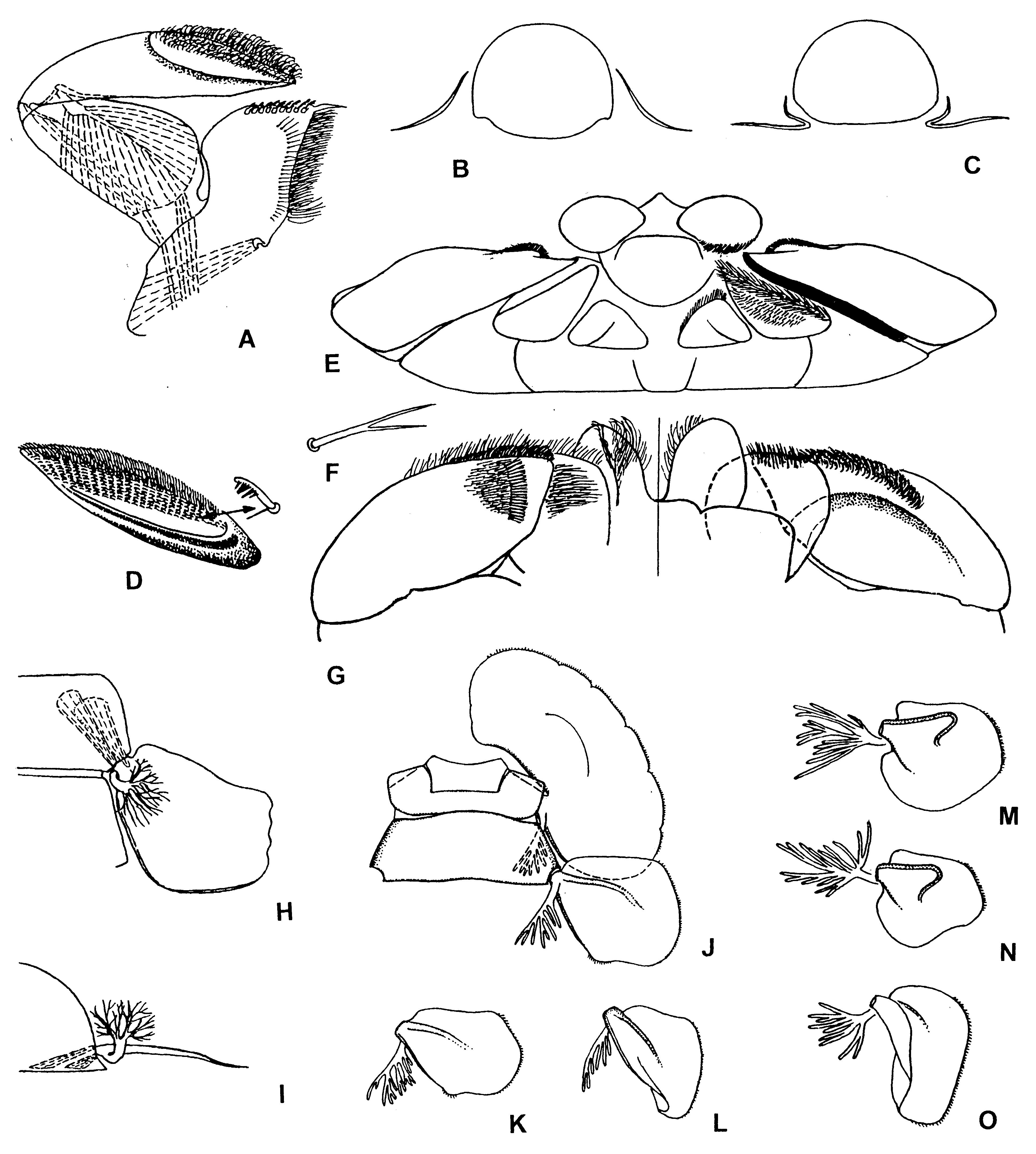
Figure 62. Rhithrogena/fg2, larvae.
A–B – Cinygmula/g1: A – right maxilla, ventral view (muscles shown by interrupted lines); B – cross section through abdominal segment VIII and tergalii VII. C–O – Rhithrogena/fg3: C – cross section through abdominal segment VIII and tergalii VII; D – apical (2nd+3rd) segment of maxillary palp and enlarged pectinate scraping seta; E – superlinguae, hypopharynx and labium, apical view; F – seta of distal row on dorsal surface of distal segment of labial palp; G – distal part of labium, dorsal view (in left half) and ventral view (in right half) (compare with Fig.57:F–H); H–I – scheme of natural position of tergalii (muscles shown by interrupted lines): H – dorsal view; I – posterior view (compare with Figs 57:C–E and 61:J–K); J–L – Rhithrogena/fg3-Sibirigena/g1* sibirica [Rhithrogena]: J – abdominal segments I and II with left tergalii, ventral view; K–L – right tergalii VI and VII, dorsal view; M–O – Rhithrogena/fg3-Epeiron/g1 znojkoi [Ecdyonurus]: right tergalii V–VII, dorsal view (fibrillose portions turned apart). (D–G – from Kluge 1988a; H–I – from Kluge 1993a; J–K – from Kluge 1997d).